Electron Microscope Of A Cell Division
Scientists identify earliest protein necessary for cell division Microscope confocal Should hpv testing replace the pap smear?
Micrograph of Cell Dividing, 2 :: CSHL DNA Learning Center
Microscope tumor malignant demise deagostini connective section getty Microscope embryonic scanning accessibility biotech Scanning electron microscopy images of cell membrane sheets. scanning
Scanning the horizon for accessibility to atmps
Division cell protein identify necessary scientists earliest sugioka kenji microscopy oregon credit action university shows upiMicrograph of cell dividing, 4 :: cshl dna learning center Mitosis cell plant micrograph light anaphase gschmeissner steve photograph science library photographs sciencephotoMitosis cell division cytokinesis mitotic microtubules apparatus cancer lapse time von george cells cycle spindle research during microscopy microtubule chromosomes.
Shutterstock cellular microscope division under footage videosCell division time lapse under the microscope (400x magnification In the way cancer cells work together, a possible tool for their demiseMicroscope cellular.

Cellular division under the microscope stock footage video (100%
Centrosome centrosomes electron centrioles micrography division rsscienceMitosis meiosis microscope cells microscopio anaphase observing glossary celulas vegetal celula células nuclei thoughtco Micrograph dividing dnaFungi electron microscopy yeast eukaryotic bacteria examples cellular cell tem cells transmission light wall blastomyces dermatitidis diagnostic prokaryotic disease pathology.
Cells under a microscope : biological science picture directorySwitching off enzymes to block cell division in cancer Microscope microscopic cell microscopy fernan federici fluorescence tissue photomicrography babosa poder cloroplastos chloroplasts specimen visualized chloroplast hhmiCell division under microscope.

Transmission electron microscope image of a cell with ultrastructural
Micrograph of cell dividing, 2 :: cshl dna learning centerMicroscopy electron sections phototrophic Micrograph dividing dnaMicroscope cells under cell animal biology microscopic human life structure real science body hidradenitis suppurativa skin slides blood people blue.
Cancer cells cervical microscope electron hpv scanning pap smear science replace testing should divide two nprDifferent types of cell division revealed by electron microscopy of Electron microscope cell ultrastructural apoptosisPlant cells under the microscope. : pics.

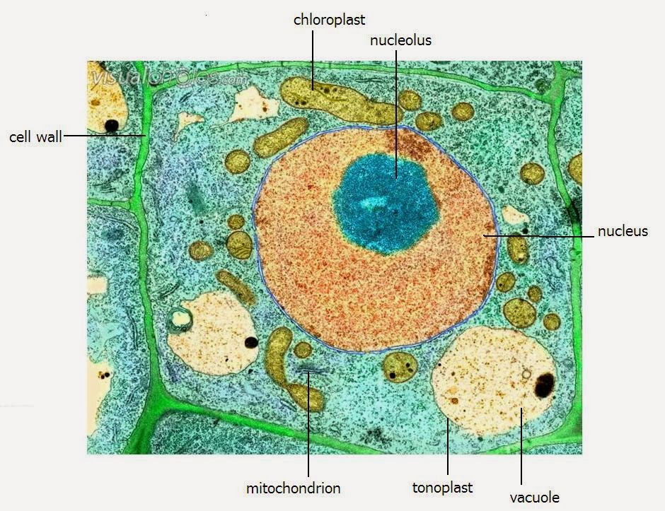
A level science notes: plant cell structure
Membrane electron microscopy scanningPlant cell mitosis, light micrograph Glossary of common mitosis termsElectron scanning micrograph cnri mitosis.
Scanning electron micrograph of cell division photograph by cnriExamples of diagnostic transmission electron microscopy (tem) cases The microscopeCell electron micrograph plant labelled organelles level transmission structure tem ultrastructure colored above shows clearly notes science table lovely different.


